Visualizing Viruses: How COXEM SEMs Are Advancing Infectious Disease Research
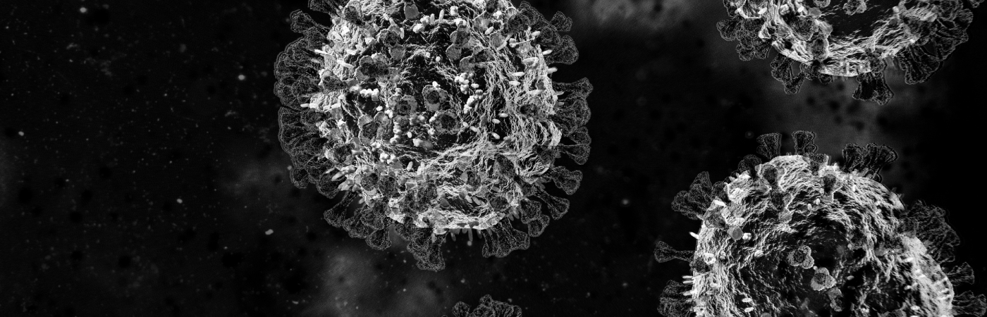
In recent years, the global spotlight has been focused on viruses like SARS-CoV-2, the causative agent of COVID-19. Understanding how such pathogens function, spread, and evolve is crucial for researchers and public health professionals alike. One of the most powerful ways to study viruses is by seeing them—literally. Scanning Electron Microscopy (SEM), particularly with compact and accessible systems like those offered by COXEM, is transforming the way virologists visualize and analyze disease-causing viruses.
While Transmission Electron Microscopes (TEMs) have traditionally been used for visualizing internal viral structures, SEMs provide detailed images of virus morphology and surface interactions. This is especially important for studying how viruses attach to and affect host cells, form viral clusters, or respond to antiviral treatments. COXEM’s benchtop SEMs, offering magnifications up to 150,000×, are proving invaluable for these purposes. Their user-friendly design and compact footprint allow researchers in university labs, biosafety level (BSL) facilities, and pharmaceutical R&D centers to gain high-resolution insights without the complexity and overhead associated with traditional electron microscopes.
One of the major advantages of using SEM in virology is the ability to observe viruses in their natural structural state—how they look on the surface, how they cluster, and how they behave when interacting with various environments. For instance, COXEM SEMs can be used to visualize SARS-CoV-2 particles adhering to human epithelial cells, or to study the budding process by which viruses exit infected cells. This type of visualization provides not only confirmation of infection but also insights into viral mechanisms of action.
Another key area where SEMs are making an impact is in vaccine development. Viral vectors—such as adenoviruses or virus-like particles (VLPs)—used in vaccines can be imaged to confirm size, uniformity, and surface characteristics. Ensuring the physical integrity of these vectors is essential for consistent immune responses. SEM images can also help researchers understand the stability of viral particles under different storage or delivery conditions, making SEM a critical part of pharmaceutical quality assurance.
COXEM SEMs are also being used to investigate airborne virus particles and contaminated surfaces—an essential application in pandemic preparedness and bioaerosol research. For example, researchers can detect virus particles captured on air filters, masks, or lab surfaces, contributing to improved infection control protocols.
Incorporating COXEM’s Energy Dispersive Spectroscopy (EDS) capabilities adds another layer of functionality, enabling elemental analysis of samples. This can be useful for tracing labeled viral particles or understanding contamination in biological environments.
For visual assets, researchers are encouraged to leverage public domain SEM images from trusted sources like the CDC Public Health Image Library, the University of Texas Medical Branch (UTMB), or platforms such as Wikimedia Commons and Pixabay. These include striking SEM images of viruses like HIV, influenza, and SARS-CoV-2 that not only enhance scientific communication but also serve as compelling visual evidence in publications and educational outreach.
In conclusion, COXEM SEMs are empowering a new generation of virologists to explore the microscopic world of pathogens with precision and clarity. By making high-resolution imaging more accessible and practical, COXEM is playing a vital role in infectious disease research, vaccine development, and public health preparedness.
If you’re working in virology, microbiology, or vaccine research and would like to see how COXEM SEMs can support your work, visit www.coxem.com or contact your regional distributor to request a demo.